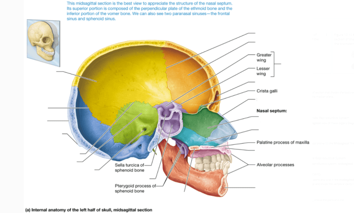
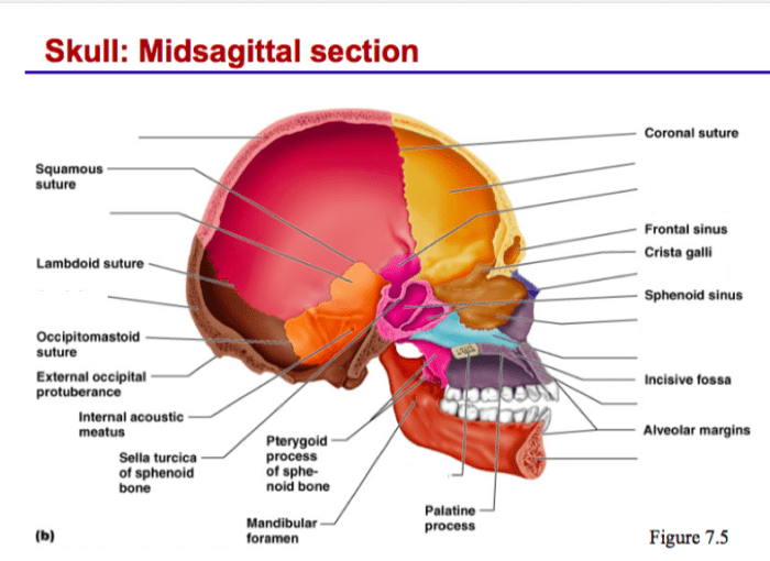
Art-labeling activity internal midsagittal view of the skull – The art-labeling activity of the internal midsagittal view of the skull offers a unique and engaging approach to understanding the complex anatomy of the skull. This activity combines artistic expression with scientific accuracy, providing students with a memorable and effective learning experience.
This detailed view of the skull’s midline reveals intricate anatomical structures, including the frontal, parietal, and occipital bones. The sagittal suture, a prominent feature in this view, plays a crucial role in the skull’s growth and development.
Internal Midsagittal View of the Skull: Art-labeling Activity Internal Midsagittal View Of The Skull

The internal midsagittal view of the skull provides a detailed look at the skull’s anatomy from the midline. This view is important for understanding the skull’s structure and function, and it is used in a variety of medical imaging techniques.
Anatomical Structures
The internal midsagittal view of the skull reveals several major anatomical structures, including:
- Frontal bone:The frontal bone forms the forehead and the roof of the eye sockets.
- Parietal bone:The parietal bones form the sides and top of the skull.
- Occipital bone:The occipital bone forms the back of the skull and the base of the skull.
- Sagittal suture:The sagittal suture is a fibrous joint that runs along the midline of the skull, connecting the frontal and parietal bones.
Clinical Significance
The internal midsagittal view of the skull is important in medical imaging because it allows doctors to visualize the skull’s anatomy and identify any abnormalities. This view is used to diagnose and treat a variety of conditions, such as:
- Skull fractures:The internal midsagittal view can be used to identify skull fractures, which are breaks in the skull bone.
- Brain tumors:The internal midsagittal view can be used to identify brain tumors, which are abnormal growths of tissue in the brain.
The internal midsagittal view is also used in neurosurgical procedures, such as surgery to remove brain tumors.
Art-Labeling Activity
An art-labeling activity can be used to help students learn and retain information about the internal midsagittal view of the skull. To complete this activity, students can use an image of the internal midsagittal view of the skull and label the different anatomical structures.
| Structure | Description |
|---|---|
| Frontal bone | Forms the forehead and the roof of the eye sockets. |
| Parietal bone | Forms the sides and top of the skull. |
| Occipital bone | Forms the back of the skull and the base of the skull. |
| Sagittal suture | A fibrous joint that runs along the midline of the skull, connecting the frontal and parietal bones. |
Educational Applications
Art-labeling activities are a valuable tool for anatomy education because they help students to:
- Visualize the anatomy:By labeling the different anatomical structures, students can create a mental image of the skull’s anatomy.
- Recall information:The act of labeling the structures helps students to remember the names and locations of the different anatomical structures.
- Apply knowledge:Art-labeling activities can be used to assess students’ understanding of the skull’s anatomy.
Art-labeling activities can be integrated into different teaching methods, such as:
- Lectures:Art-labeling activities can be used to supplement lectures on the skull’s anatomy.
- Labs:Art-labeling activities can be used in labs to help students identify the different anatomical structures of the skull.
- Independent study:Art-labeling activities can be assigned as homework to help students review the skull’s anatomy.
Extensions, Art-labeling activity internal midsagittal view of the skull
There are several ways to extend the art-labeling activity, such as:
- Using different materials:Students can use different materials to label the anatomical structures, such as markers, crayons, or paint.
- Creating a 3D model:Students can create a 3D model of the skull and label the different anatomical structures.
The art-labeling activity can also be adapted for different age groups and learning levels. For example, younger students can be given a simplified image of the skull to label, while older students can be given a more detailed image.
FAQ Resource
What is the purpose of the art-labeling activity?
The art-labeling activity aims to enhance anatomical knowledge and understanding of the internal midsagittal view of the skull through artistic expression and labeling.
How does the activity contribute to learning?
By actively engaging with the material and labeling different anatomical structures, students reinforce their understanding of the skull’s anatomy and its clinical significance.
What are the benefits of using an art-labeling activity in anatomy education?
Art-labeling activities foster creativity, critical thinking, and a deeper understanding of anatomical structures and their relationships.

